
Understanding Usher syndrome can feel overwhelming at first, especially when you’re trying to make sense of new medical terms. This section explains the condition clearly and simply, with trusted information created with experts.
What is Usher Syndrome?
Usher syndrome is a rare genetic condition which causes hearing loss, progressive vision loss, and, in some cases, balance difficulties (vestibular dysfunction). Vision loss in Usher syndrome is caused by retinitis pigmentosa (RP), which affects the area at the back of the eye which senses light (the retina). The first symptom is usually difficulty seeing in the dark, followed by progressive loss of peripheral vision (reduced visual field).
Usher syndrome is thought to affect between 4-17 individuals per 100,000 in the population. We estimate there are approximately 2,000 children and young people aged 0-25yrs with Usher syndrome living in the UK.
This video, produced in partnership with Retina UK, gives basic information about Usher syndrome. This may be helpful in providing an understanding of the condition to wider family members or professionals supporting a child with Usher syndrome.
Our newly diagnosed information pack (PDF) is another useful resource giving an overview of Usher syndrome.
If you’d like more detailed information you can access the sections using the menu on this page to find out more about the different aspects of Usher syndrome.
What are the types of Usher syndrome?
There are 4 main clinical types of Usher syndrome, each associated with different genetic changes.
| Type | Hearing loss | Vision loss | Balance difficulties |
|---|---|---|---|
| Type 1 | From birth – severe to profound | Usually affects individual by age 10 to 20 | From birth |
| Type 2 | From birth – moderate to severe | Usually affects individual by age 20 to 30 | Usually not affected |
| Type 3 | Progressive – moderate to severe | Variable | Variable |
| Type 4 | Progressive – beginning in adulthood | Usually affects individual by age 50 to 60 | No issues |
The age and rate at which changes to vision take place varies between individuals, even with the same type of Usher syndrome.
How is Usher syndrome diagnosed?
To confirm a diagnosis of Usher syndrome, full genetic testing and ophthalmology tests are needed.
Genetic testing should be offered once hearing loss is identified (for example, if a baby is diagnosed with hearing loss by an audiologist following newborn hearing screening) or can be requested via the audiologist or paediatrician (if involved), if it has not been offered.
For the genetics test, a blood sample will be taken from the child and both parents, and sent for analysis (which can take several months), leading to a full genetic report. A Genetics Counsellor can help explain the information within the full genetic report, and arrange referrals if required (for example to Ophthalmology).
Ophthalmology tests carried out in a hospital eye clinic will include electro-diagnostic tests (an electroretinogram, ERG) and a consultation with the Ophthalmologist. They will review the results of the tests alongside the genetic report and other relevant symptoms, in order to confirm the diagnosis of Usher syndrome.
You can find more information about the tests involved in diagnosis in our newly diagnosed information pack (PDF) (opens in a new tab).
How is hearing affected by Usher syndrome?
Structure and function of the ear
In this section we will give information about the structure and function of the ears, and how hearing is affected by Usher syndrome.
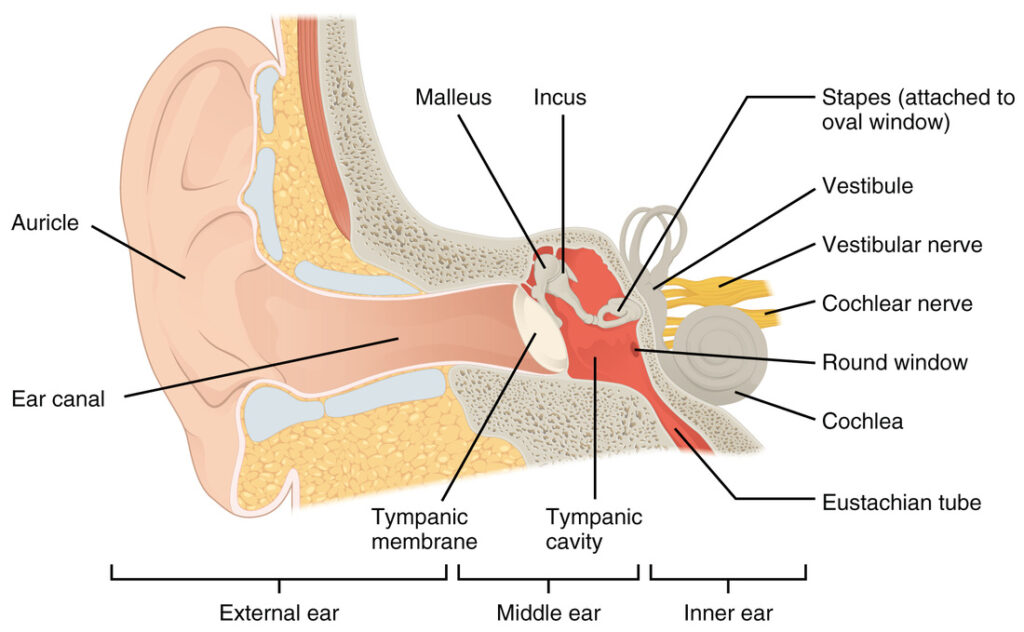
Structure
Outer Ear
- Auricle: This is the outer part of the ear situated on either side of the head.
- Ear canal: This canal is lined by skin and passes from the hole in the auricle through cartilage and bone to the tympanic membrane (ear drum).
- Tympanic membrane: This is the thin transparent piece of skin which sits at the end of the ear canal
Middle Ear
- This is a small, air-filled, bony chamber, that contains three small bones connecting the tympanic membrane to the inner ear.
Inner Ear
This consists of two parts:
- Cochlea: This spiral shaped, fluid-filled structure has an elastic membrane along its entire length. Thousands of hair cells are aligned in 4 rows on top of the membrane. They are activated when the hairs on their upper end bend with resonating vibration. This “bending” is limited to the places where sound is resonating with the membrane. Each of the thousands of hair cells is connected to its own nerve and the brain is able to differentiate sounds based on where and which hair cells are triggered from the membrane’s resonance. This association is called “tonotopic coding of sound”.
- Semi circular canals: These 3 interconnecting tubes are also filled with fluid.
- Vestibulo-cochlear nerve: This nerve is connected to both the cochlea and the semi circular canals. Each nerve fibre in the cochlear part of this nerve is attached to just one hair cell. This nerve conducts messages to the hearing centre in the brain.
Function
When a sound is present it sets up vibrations in the ear which pass through the ear canal to the tympanic membrane and the three small bones in the middle ear. Together these structures increase the force of the vibrations entering the cochlea. The hair cells bend with this resonating vibration and stimulate its attached nerve fibre. The nerve fibres then pass information about the sound to the part of the brain that deals with hearing.
The Usher Syndrome ear
The affected genes that cause Usher Syndrome are also responsible for a number of proteins that are found in the hair cells of the inner ear. These proteins are required to ensure healthy growth of the hair cells. The hair cells are called stereocilia and are responsible for converting the vibrational waves of sound into the electrical signals sent to the brain. The lack of these proteins causes the majority of the stereocilia to be underdeveloped and non functional, therefore unable to convert the sound to electrical signals. When these electrical signals aren’t able to get through to the auditory nerve and then onto the brain, it results in deafness in Usher syndrome patients.
How is hearing tested in children?
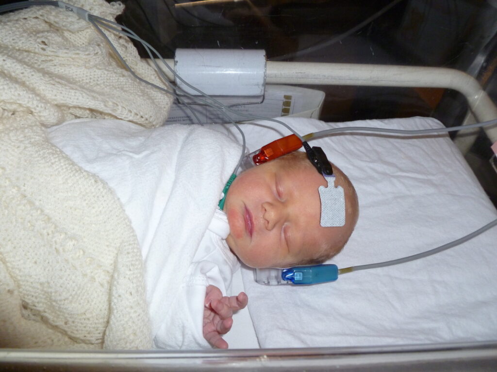
Newborn Infant Hearing Screening
All areas in the UK have universal neonatal hearing screening programmes. These programmes screen the hearing of all newborn babies in their first few weeks of life. They aim to find out as early as possible whether a baby has a hearing loss. The screen is performed by trained hearing screeners using valid and reliable technology and is usually carried out at the baby’s bedside in hospital while they are asleep.
Auditory Brainstem Response (ABR)
The ABR is the most common test given to babies during the screen. Sensors are placed on the baby’s forehead and behind the ears with some sticky gel (see image above). Clicking sounds are then played through an earpiece placed inside the baby’s ear while they are asleep. Brain responses to these sounds are recorded. These brain responses provide information about the softest level of sound the baby can hear. This information can help confirm whether further testing or further interventions (such as hearing aids) or special education may be recommended.
Audiogram
For older children, behavioural tests are used to determine a child’s response to sound. Each ear is tested separately with the ear not being tested covered with a soundproof earpiece. A series of sounds are produced by a machine and relayed to the ear being tested. The sounds vary according to their pitch or frequency (cycles/second) and loudness (decibel level). The loudness of the sound detected by the child is recorded for several frequency levels between 250 and 8000 cycles per second (see below images).
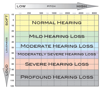
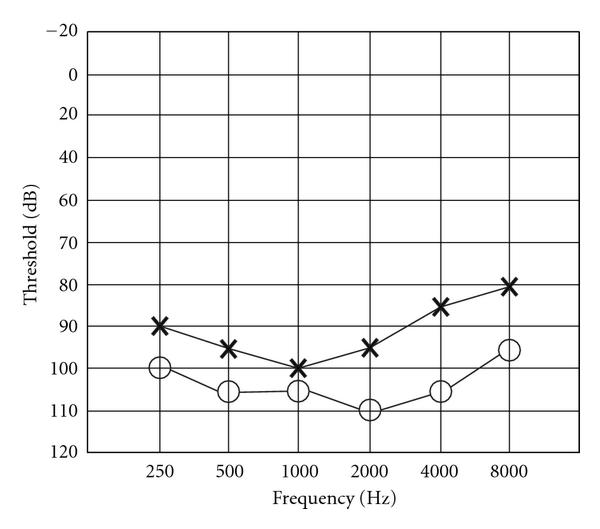
How can hearing loss be managed?
It is important to understand that each child has different needs when it comes to communication and these needs may change over time. The right approach to communication is one that works best for the individual child and their family. In consultation with a range of different support agencies and specialists, families can make informed decisions about the communication needs of their child.
Progressive audiological assessment
Neonatal screening for hearing loss takes place in all areas of the UK. Tests to monitor the progress of any detected hearing loss will be necessary to determine the appropriate time to provide further intervention.
Early fitting of hearing aids
Consultation with an ENT surgeon and audiologist will be important when deciding whether your child will benefit from using hearing aids. Even though hearing aids will not provide enough sound for some children to acquire speech due to their degree of hearing loss, at the very least evidence suggests that early fitting of hearing aids helps to keep the auditory nerve healthy while investigating other intervention options.
Cochlear implant
For some children an implant may be an option. In this situation, special tests of hearing, consultation with an ENT surgeon and consideration of other social and personal factors will be important. Implantation as early as six to twelve months gives best results.
Sign language
British Sign Language is the sign language of the British Deaf community. It can be accessed through many Early Intervention and Education settings across the UK. It can be learnt in conjunction with, or exclusive of, assisted hearing devices.
How is vision affected by Usher syndrome?
Structure and function of the eye
In this section we will give information about the structure and function of the eyes, and how vision is affected by Usher syndrome.
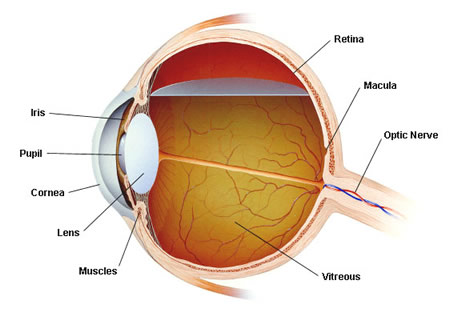
The eye consists of several parts that are somewhat similar to a camera:
- Sclera: the white material which we normally see as the white of the eye. It is the eye‘s outer protective coat.
- Cornea: the transparent, curved structure at the front of the eye
- Iris: through the cornea the iris can be seen, which is that part of the eye which gives it colour – blue, brown, green, grey etc. It is a circular muscle which responds to bright light by contracting, making the pupil smaller.
- Pupil: in the middle of the iris is the pupil, a transparent aperture which appears black. The pupil constricts in response to bright light and dilates in low levels of light.
- Lens: situated immediately behind the iris and pupil is a transparent disc with both sides being convex. The lens focuses light onto the retina. It divides the eye into two parts – anterior and posterior chambers.
- Anterior Chamber: a fluid with the consistency of water circulates around this chamber. This fluid is called the aqueous humour.
- Posterior Chamber: this is filled by the vitreous humour which has the appearance of transparent jelly.
- Retina: situated at the back of the eye, the retina consists of millions of nerve cells which are divided into two main groups – rods and cones. They are so described because of their appearance under a microscope.
- Cones: concentrated around an area of the retina called the macula.
- Rods: although some are placed near the macula, the majority of rods are spread out to cover the rest of the retina.
- Retinal Pigment Epithelium: a dark coloured layer of cells underlying the retina responsible for providing oxygen and other nutrients to the rods and cones.
- Choroid: located behind the retina, it consists of a large network of blood vessels that transport oxygen and other nutrients to the retinal pigment cells.
- Optic Disc: the nerve cell connections from all the rods and cones travel to the optic disc, a small, yellow, oval structure which can be seen through an ophthalmoscope.
- Optic Nerve and beyond: the optic disc is the front part of the optic nerve which passes from the eye to make connections throughout the brain.
How vision is affected by Usher syndrome
Vision loss in Usher syndrome is caused by retinitis pigmentosa (RP), which affects the area at the back of the eye which senses light (the retina). The first symptom is usually difficulty seeing in the dark, followed by progressive loss of peripheral vision (reduced visual field).
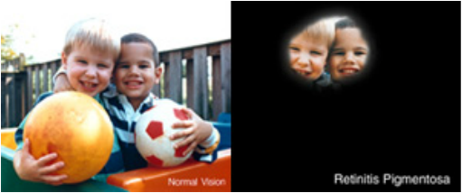
Retinitis Pigmentosa
Retinitis Pigmentosa, also known as RP, is an inherited eye condition affecting the retina light-sensitive tissue at the back of the eye (retina). When optometrists or ophthalmologists use an ophthalmoscope to look into the eye of a person with RP, they usually see scattered areas of pigment across the retina. By contrast, in a normally sighted person, the pigmented cells are covered by the living nerve cells – the rods and cones. The pigmented appearance gives rise to the name – retinitis pigmentosa.
People affected with RP have difficulties with seeing at night, loss of peripheral (side) vision and may have a tendency to trip over curbs and stairs. Symptoms and rate of visual loss can vary between individuals. At present there is no treatment or cure, but many research groups both in UK and overseas are looking at potential future therapies. Although there is no current treatment for Usher syndrome, it is important to maintain eye checks as cataracts and macula odema (swelling) can be associated with RP and are able to be managed.
When do symptoms of RP begin?
Individuals diagnosed with Usher syndrome type 1 will usually start to develop symptoms of RP within the first decade of their life. Those with Usher syndrome type 2, the symptoms usually appear in the second decade of life.
The rate of vision loss is usually slow, this is why the diagnosis without genetic testing is often not made until the child is in their early teens.
How can vision loss be managed?
There is currently no treatment for Usher syndrome. Knowing the diagnosis means that monitoring and support can be put in place, and skills can be developed in anticipation of potential future vision loss. It also means that researchers can learn more about the condition in order to develop future treatments.
The type of vision loss in Usher syndrome is called Retinitis Pigmentosa. It is not corrected by the use of glasses or other tools. However, regular testing (usually yearly) at an eye clinic is important to monitor any changes to vision, and also check for any other treatable eye conditions which are not connected to Usher syndrome but may affect vision (e.g. changes in visual acuity, or cataracts).
The tests carried out at the eye clinic will include Optical Coherence Tomography (OCT) and Fundus Autofluorescence Imaging (AF), along with checks by an Ophthalmologist. For children able to carry out visual field tests, these will also be included in annual checks.
Between visits to the eye clinic, it is really helpful if the child (or their family) can keep track of any changes they experience in their vision (for example, things which have become more difficult to see or do) to discuss with the professionals at the eye clinic. This information helps the professionals to get a more accurate picture of the child’s functional vision, which isn’t always easy to find out through tests.
If you don’t understand the information or test results being shared at the appointment please do ask the professionals carrying out the tests. It is important that you (and your child as they get older) understand this information, and feel confident to ask questions.
Many young people find assistive technology and the accessibility tools available on mainstream devices help them in their daily lives, and some people find that specialist low vision tools can be helpful for specific tasks (these can be accessed via the low vision clinic at the eye hospital).
Finally, experts recommend eating a healthy diet consisting of fresh brightly-coloured fruits and vegetables (including green leafy vegetables), alongside the use of UV protected sunglasses, in order to maximise eye health.
How is vision tested in children?
There are many procedures used to test different visual functions. Some tests can be carried out by professionals such as nurses, special education teachers or general practitioners, while others may require more specialist knowledge or equipment such that an optometrist or ophthalmologist may possess.
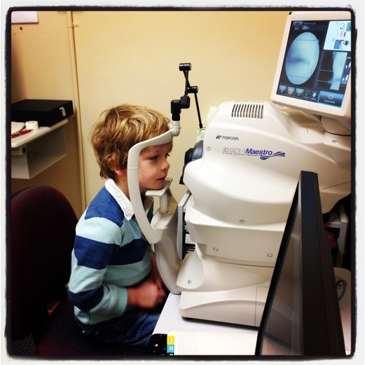
Optical coherence tomography (OCT) is a non-invasive imaging test that uses light waves to take cross-section pictures of your retina, the light-sensitive tissue lining the back of the eye.
Visual Acuity
Visual Acuity measures the ability to read the letters on an eye chart. It is the most common clinical measurement of visual function. The standard definition of normal vision is 6/6 (metric) or 20/20 (imperial). In young children vision is measured using symbols and pictures. Children who can’t understand these tests are shown different sized stripes and their behaviour is observed.
Visual Fields
A Visual Field test measures the field of vision (side vision). This is where you look straight ahead at a particular point in a bowl-shaped machine. Each time you spot dots of light you click a button. This is referred to as computerized visual field testing. Alternatively, it can be performed manually whereby a light is brought in from the side of the machine and slowly moved to the center of vision. You press a button as soon as you see the light. The results are then marked on a chart and compared with the results a person with normal vision would be expected to see. A normal visual field is described as being about 140 degrees in every direction. Assessment of visual fields becomes relevant around 8 years of age.
Colour Vision
Colour vision can be assessed by different methods, but the most commonly used test is the Ishihara Colour Vision Test. The Ishihara is a booklet that shows numbers composed of different coloured dots. The numbers are printed within different coloured dotty backgrounds.
Electroretinogram (ERG)
The ERG is used to measure the function of the retina and identify layers of the retina that are not working properly. In some cases it can detect very early signs of RP. It involves placing special electrodes either in the eye or close to the eye. Flashes of light are used to stimulate the retina and the electrodes measure the electrical response of the retinal cells to the flashing lights.
Optical Coherence Tomography (OCT)
The OCT takes cross- sectional images of the retina. It can measure the thickness of the retina and detect any retinal abnormalities such as macula odema which can occur in patients with RP.
Fundus photographs
Using special cameras, photographs are taken of the retina to document RP related eye changes.

How is balance affected by Usher syndrome?
People with Usher syndrome type 1 and approximately half of those with Usher type 3 experience difficulties with balancing, due to dysfunction of the vestibular system. Children affected by vestibular dysfunction may reach the typical motor milestones later than their peers. They may find it hard to sit upright around 6 months of age, or may have limited neck or head control. The child may appear clumsy and more prone to falls or injury. Whilst these children typically take longer to walk independently than their peers without vestibular dysfunction, once they reach these motor milestones there is usually no stopping them, just like any other child!
Here are two short videos, created in partnership with Great Ormond Street Hospital, that explain more about the vestibular system, and how it is affected by Usher syndrome.
How is balance affected by Usher syndrome?
How does vestibular function work and how is it tested?
How does the balance system work?
The body’s balance system works through a constant process of position detection, feedback and adjustment using communication between the inner ear, eyes, muscles, joints and the brain.
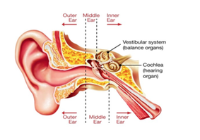
Deep inside the ear, positioned just under the brain, is the inner ear. While one part of the inner ear enables hearing, another part, called the vestibular system, is designed to send information about the position of the head to the brain’s movement control centre, the cerebellum.
The brain’s movement control centre
The cerebellum is a small part of the brain positioned at the back of the head, where it meets the spine, which acts as the body’s movement and balance control centre. It receives messages about the body’s position from the inner ear, eyes, muscles and joints, and sends messages to the muscles to make any postural adjustments required to maintain balance. It also coordinates the timing and force of muscle movements initiated by other parts of the brain.
Position feedback from the inner ear
The vestibular system in each inner ear is made up of three semi-circular canals and two pockets, called the otolith organs, which together provide constant feedback to the cerebellum about head movement.
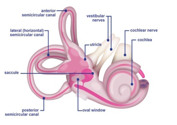
Each semi-circular canal has a different orientation to detect a variety of movements such as nodding or rotating. Movement of fluid inside the canals caused by head movement stimulates tiny hairs that send messages via the vestibular nerve to the cerebellum.
The two otolith organs (called the saccule and utricle) send messages to the brain about body movement in a straight line (backwards/forwards or upwards/downwards) and also about where the head is in relation to gravity, such as tilting, leaning or lying down. These organs contain small crystals that are displaced during these movements to stimulate tiny hairs, which transmit the message via the vestibular, or balance nerve to the cerebellum.
Position feedback from eyes, skin, muscles and joints
The vestibular system (inner ear balance mechanism) works with the visual system (eyes and the muscles and parts of the brain that work together to let us ‘see’) to stop objects blurring when the head moves. It also helps us maintain awareness of positioning when, for example, walking, running or riding in a vehicle. In addition, sensors in the skin, joints and muscles provide information to the brain on movement, the position of parts of the body in relation to each other, and the position of the body in relation to the environment. Using this feedback, the brain sends messages to instruct muscles to move and make the adjustments to body position that will maintain balance and coordination.
If any of these systems isn’t working right, it can affect balance.
Reprinted with permission from Royal Victorian Eye and Ear Hospital.
How can the balance effects be managed?
Children with vestibular dysfunction will often be delayed in reaching motor milestones, but they do get there! In the early years they may demonstrate reduced head control, delayed independent sitting, and/or delayed walking. The child may continue to appear clumsy and more prone to falls or injury. As they develop other skills and strategies to compensate for the vestibular dysfunction, the everyday impact can be minimised. The videos below cover some strategies and exercises which can be used to support balance development in children with vestibular dysfunction caused by Usher syndrome.
Very little specialist support or advice is available on what strategies and exercises can help support balance, but the general advice has always been to encourage the child to be as physically active as possible, and also for them to explore surfaces with bare feet as often as possible. Being physically active and exploring surfaces using touch will allow them to develop the proprioception responses which will compensate for lack of balance function. Below are two short films created in partnership with Great Ormond Street Hospital:
What strategies can help with balance?
What exercises support the vestibular system?
Vestibular rehabilitation therapy (VRT) – is an exercise-based program run by specialist Vestibular Physiotherapists and is designed to promote central nervous system compensation for inner ear deficits. VRT exercises should be personalised based on a child’s age, interests, level of comprehension, and test results. The exercises may help strengthen balance skills and coordination. Exercises might involve things like bending down, standing or walking with eyes open and then with eyes closed, swimming, or walking barefoot on various uneven surfaces.
Children typically respond more quickly to VRT than adults, because of their greater neuroplasticity—the ability of their neurological systems to more quickly compensate for and adapt to vestibular deficits. In addition, children tend to be less fearful of movement than adults, so they participate well in the balance and movement aspects of therapy.
Safety Considerations
While they are working on their balance, it is important to make sure all those who look after the child know strategies to keep them safe (e.g. extra cushions to protect them if sitting unaided).
In particular, it is very important for everyone to be aware that a child without vestibular function will be unable to detect ‘which way is up’ if they are underwater. A simple strategy called the ‘mushroom float technique’ can be taught to help them reach the surface of the water (online videos available to help teach this skill), and in the meantime they need close support and supervision whenever they are in water.
How common is Usher syndrome?
Experts think that Usher syndrome affects between 4-17 people in every 100,000 people. In the UK, it is thought that approximately 2,000 children and young people aged 0-25yrs live with Usher syndrome, but not all of these will have been diagnosed.
What are the chances of inheriting Usher syndrome?
Genes are the instructions that our bodies need to grow and develop. There are a number of genes known to cause Usher syndrome and there are likely to be additional genes still undiscovered. These are genes that have a role in the inner ear and the retina.
Each of these genes are found in pairs, with one inherited from each parent. Usher syndrome is inherited in an autosomal recessive way. This means that both copies of the gene (the one from mum and the one from dad) need to not be working to cause Usher syndrome. Usually this means that mum and dad are each a “carrier” for the condition, having one working copy and one non-working copy. Carriers do not have the condition. There is nothing anyone did or did not do to cause themselves to be a carrier or their child to have Usher syndrome.
When two carriers of Usher syndrome have children together, each time there is a:
- 1 in 4 (25%) chance that a child will have two working copies of the Usher gene. This child does not have Usher syndrome.
- 2 in 4 (50%) chance that a child will have one working and one non-working copy of the Usher gene. This child is a carrier like their parents.
- 1 in 4 (25%) chance that a child with have two non-working copies of the Usher gene. This child has Usher syndrome.
This is the same for male and female children. This is a random process, so the % chances listed above apply to each new pregnancy.
The Gene Vision website is an excellent source of detailed information about genetic eye conditions.
Stay in touch
We send out emails to keep people on our mailing list up to date with events, new resources and other services which we think are relevant to our community.